Is the ITB Connected to the Lateral Meniscus?
You would think that in the early 21st century we would have figured out everything there is to know about the human knee, but you would be wrong. In fact, it was just a few years ago that the anterior lateral ligament was discovered. How is that possible? Because physicians and anatomists have a hard time imagining anything other than what they have been taught. So today I’ll announce another possible discovery, that the ITB and the lateral meniscus are connected.
Hiding in Plain Sight
It’s often hard to conceptualize something differently from what you’ve been taught. Take, for example, this picture of Leonardo da Vinci’s last supper:
You clearly see Jesus in the middle. That woman to the left? That was assumed for centuries to be a man! There’s no doubt to anyone who wasn’t educated in art history that it’s clearly a woman. To art historians, that position to the left of Jesus (as we look at the painting) is always occupied by the apostle John. Hence, despite the fact that all the men are shown with beards or square jaws, this female figure must be John.
Discovering the ALL Ligament
You would think that all discoveries of the anatomy of the human knee happened well over a hundred or more years ago. However, nothing could be further from the truth. In fact, it was only in the 1970s when physicians believed that the meniscus was just an embryologic remnant and unimportant rather than a living and critical stabilizer and cushion protecting cartilage. Then, just a few years back, anatomists discovered a new ligament in the knee called the ALL (anterior lateral ligament). This ligament on the outside of the knee mimics the ACL (anterior cruciate ligament) function and is thought to be one reason why ACL reconstruction surgery is less than 100% successful at restoring normal knee biomechanics. So we are still discovering stuff about the knee.
What Is the ITB?
The iliotibial band (ITB) is a fibrous tissue band that extends from the ilium (side of the hip) to the tibia (leg bone) on the outside of the thigh. For part of its run, it’s actually the lateral wall of the vastus lateralis muscle. See my video below to learn more:
What Is the Meniscus?
The meniscus is the figure-8 shaped fibrocartilage cushion between the knee joint surfaces. It’s a living piece of tissue that both helps to protect the joint surfaces and acts as a stabilizer for the joint. It has many things that connect to it from outside the knee, including a portion of the hamstrings tendon as well as the popliteus muscle.
What Is Tensegrity?
Tensegrity is how your body functions to passively stay upright and stable. At its simplest, it’s structural pieces that are under tension via connections that are flexible. To understand what this looks like as a model, see the video below:
Hiding in Plain Sight on MRI Images
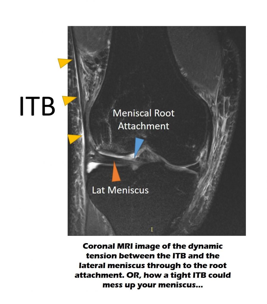
So hidden things often hide in plain sight, and everyone from physicians to anatomists conceptualize it in the context of what they have been taught, even if that means ignoring what they clearly see. Take, for example, this MRI image that I see all the time:

What do you see? The dark line coming down the left-hand side of the image and that’s being denoted by the triangle pointers is the ITB. The lateral meniscus is shown as the thicker dark structure that goes from left to right. The interesting thing is that both of these structures are clearly connected. This connection isn’t commonly seen, but here the knee is swollen, so the bright fluid outlines the attachment.
To me, what I have shown you here has always represented a connection between the lateral meniscus and structures outside the knee that balance the tension of the meniscus. Meaning that this is a tensegrity system whereby the outside forces pull one way and the attachment of the meniscus to the bone through its root pulls the other way. However, what’s bizarre is that nowhere is this connection described in anatomy texts or in papers that describe the meniscus.
The Push Back
I posted the above image on LinkedIn and immediately got push back from physical therapists who correctly noted that there is no published literature on this connection. Why would this connection be a big deal? The ITB travels from the hip to the knee, and its muscles are commanded by spinal nerves that come from the low back. So this would be yet another tie in between the low back, hips, and knee.
I then looked at my own ITB and meniscus and took this video of me contracting my ITB:
As you can see, if you look at the structure I have labeled as “Men,” which is my lateral meniscus, by contracting my ITB I can make the meniscus move. Which could suggest a connection between the two structures? Is this concrete 100% proof of a connection? Not yet.
The Next Steps
I recently performed hydrodissection on this area with an injection of fluid using ultrasound. This injection of fluid around this area wasn’t able to ply apart the meniscus and the ITB. This again suggests that the two are connected.
Next, we’ll physically dissect out one of the cadaver knees from one of the many IOF courses taught at our facility. This should confirm or deny that this connection exists. Even if it can be found in some patients, that doesn’t mean that we’ll see it in all patients as there are many anatomical variations out there.
Why This and Other Connections of the Knee Meniscus Are a Big Deal
The reason why all of this matters is that the more meniscus connections there are between muscles that go from the pelvis to the knee, the more the two are related. Meaning, if an irritated low back nerve causes some of these muscles to misfire, that can chew up the knee meniscus. All of that matters, as we harm many patients because physicians often don’t understand how the body is connected. Importantly, doctors love to divide the body into this part or that part. If interventional orthopedics is going to replace surgical orthopedics, an understanding of how everything is connected is critical.
The upshot? The hip bone is connected to the knee bone. Figuring out how that all works is critical to help understand how best to help patients with knee problems.

If you have questions or comments about this blog post, please email us at [email protected]
NOTE: This blog post provides general information to help the reader better understand regenerative medicine, musculoskeletal health, and related subjects. All content provided in this blog, website, or any linked materials, including text, graphics, images, patient profiles, outcomes, and information, are not intended and should not be considered or used as a substitute for medical advice, diagnosis, or treatment. Please always consult with a professional and certified healthcare provider to discuss if a treatment is right for you.